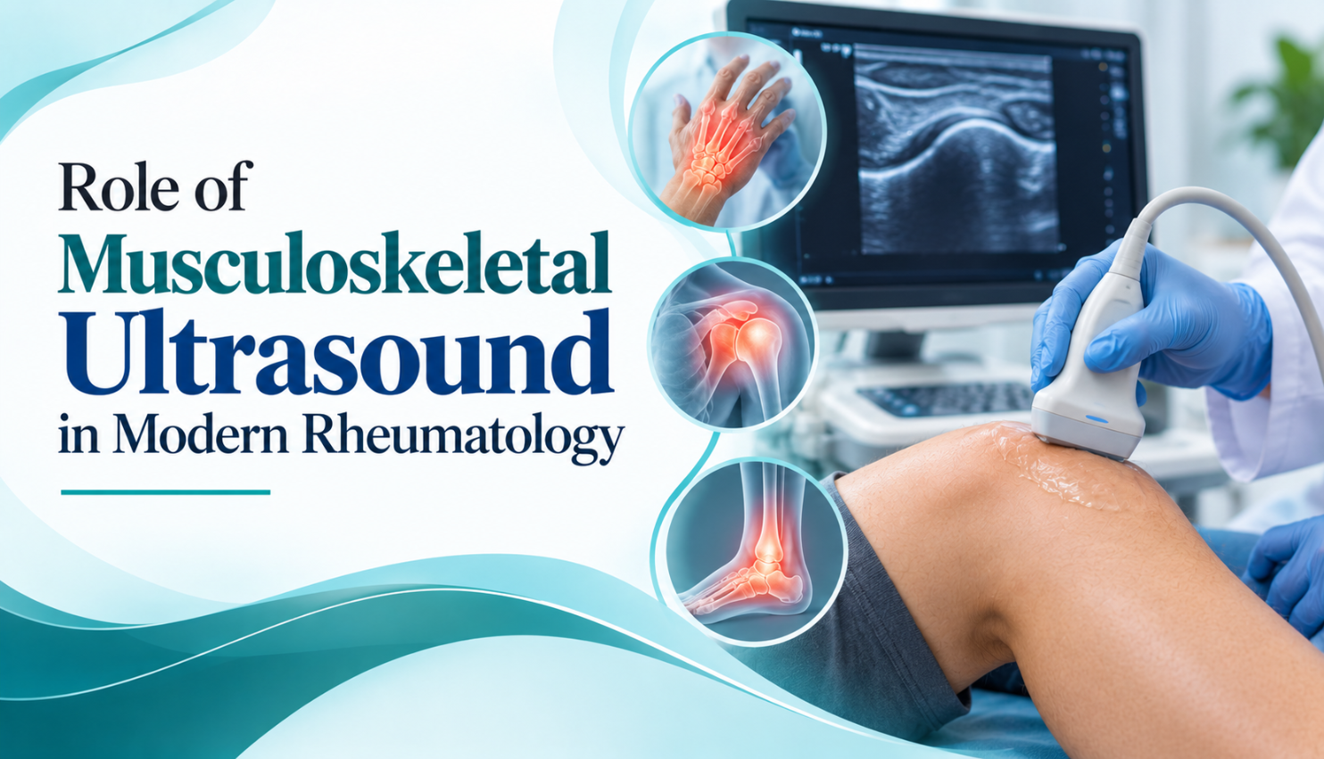
Musculoskeletal ultrasound has transformed how rheumatologists diagnose and monitor conditions like arthritis, gout and tendon injuries. This blog explains how Dr. Sandeep Nagar, a EULAR-certified specialist, uses this radiation-free, real-time imaging technique to detect early inflammation, guide precise joint injections and track treatment response over time. Learn why this advanced diagnostic tool allows faster, more accurate care during a single consultation, and how it supports better, more consistent long-term outcomes for patients with autoimmune and musculoskeletal conditions.
Musculoskeletal ultrasound has become one of the most valuable tools in modern rheumatology, transforming the way conditions such as rheumatoid arthritis, gout, tendinitis and soft tissue injuries are diagnosed and monitored. Unlike X-rays, which primarily show bone, ultrasound provides a real-time, dynamic view of joints, tendons, ligaments and the synovial lining, allowing a trained rheumatologist to detect inflammation and early damage often long before it would appear on conventional imaging. Dr. Sandeep Nagar, having completed specialised musculoskeletal ultrasound training and holding a EULAR certification in the technique, uses this skill routinely to support accurate, timely diagnosis for his patients.
One of the key advantages of musculoskeletal ultrasound is its ability to detect synovitis, or joint lining inflammation, at a very early stage. In conditions like rheumatoid arthritis, subtle synovial thickening and increased blood flow, visible through a technique called power Doppler imaging, can indicate active disease even when a joint appears normal on physical examination alone. This early detection allows treatment to begin sooner, which is directly linked to better long-term outcomes, since early, effective control of inflammation helps prevent irreversible joint damage down the line.
Musculoskeletal ultrasound is also extremely useful for guiding minimally invasive procedures. When a joint injection is required, whether to relieve pain, reduce inflammation or confirm a diagnosis through fluid analysis, ultrasound guidance allows the needle to be placed with far greater precision than a landmark-based approach alone. This improves both the safety and effectiveness of the procedure, reduces discomfort for the patient, and increases the likelihood that the treatment will provide meaningful, lasting relief rather than only partial or short-term improvement.
Beyond rheumatoid arthritis, musculoskeletal ultrasound plays an important role in diagnosing and monitoring a wide range of conditions Dr. Nagar treats regularly. In gout, ultrasound can identify characteristic urate crystal deposits within joints, helping distinguish gout from other forms of arthritis without always requiring joint fluid aspiration. In tendinitis, bursitis and soft tissue injuries, it clearly visualises inflammation, tears or fluid collections, allowing for more targeted, effective treatment planning. It is also useful in assessing enthesitis, or inflammation where tendons attach to bone, a hallmark feature of spondyloarthritis.
For patients, one of the most reassuring aspects of musculoskeletal ultrasound is that it is painless, radiation-free and can typically be performed during the same consultation, without the wait times associated with referral to a separate radiology centre. This means Dr. Nagar can often confirm a diagnosis and begin discussing treatment options within a single visit, rather than requiring patients to return for a follow-up appointment once external imaging results become available, saving valuable time during periods of active symptoms.
Ultrasound is also proving increasingly valuable for monitoring treatment response over time. Rather than relying solely on how a patient feels, which can be influenced by pain tolerance and day-to-day variation, repeat ultrasound assessments provide objective evidence of whether inflammation is genuinely settling with treatment. This is particularly useful in patients who appear to be improving clinically but may still have subclinical synovitis, helping guide decisions about whether current treatment should continue, be adjusted, or be considered for eventual tapering.
Dr. Nagar’s use of musculoskeletal ultrasound reflects a broader philosophy of combining advanced diagnostic tools with attentive, personalised clinical care. Rather than depending on a single test or symptom description, this approach allows for a fuller, more accurate picture of what is happening within the joint, supporting treatment decisions that are genuinely evidence-based rather than assumption-driven, and ultimately helping patients achieve better, more consistent long-term outcomes in the management of their condition.
If you are living with joint pain, swelling or a suspected autoimmune condition, Dr. Sandeep Nagar, Consultant Rheumatologist , offers ultrasound-supported evaluation for precise, timely diagnosis. Book a consultation to benefit from this advanced, patient-friendly approach to rheumatology care.
As musculoskeletal ultrasound technology continues to improve, its role in everyday rheumatology practice is only expected to grow. Higher-resolution probes and refined Doppler techniques now allow even finer detail to be captured, further improving diagnostic confidence. For patients, this steady progress translates into earlier detection, more precisely targeted treatment and, ultimately, a greater chance of preserving joint function and quality of life over the course of a long-term autoimmune or musculoskeletal condition.

Dr. Sandeep Nagar is a board-certified rheumatologist practising as Consultant Rheumatologist at Yatharth Superspeciality Hospital, Greater Noida. With training from AIIMS, New Delhi, IMS and SUM, Odisha, and international certifications from MRCP, UK and EULAR, Switzerland, he offers expert, compassionate care for rheumatoid arthritis, lupus, spondyloarthritis, gout and other autoimmune conditions. Book a consultation today for personalised rheumatology care you can trust.
